|
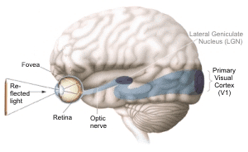
| Signals
about particular elements of the
image (of the pencil in our |
| example)
then are transported to selected
areas of the Primary |
| Visual
Cortex, or the V1,
of the brain. From there, the
signals are |
| sent
to "higher" areas of
the cortex that process more
general |
| aspects
of the image (of the pencil)
such as its shape, colour, or |
| motion
(or no motion as virtually in
our case). |
| The
various parts of the retina are so
to say 'represented' in
these |
| areas of the brain. The part that
corresponds with the fovea
centralis |
| -the
area of the retina perpendicular
behind the eye lens where |
| vision
is best- is equal in size with
the area that corresponds with
the |
| other
parts of the retina. This part
is divided in two sub areas: |
| a
sensor visual central area,
where colour, size, form, motion
and |
| clearness
are perceived, and a psycho visual
area that almost |
| surrounds
it (except in the front) where
identification and spatial |
| valuation
of the perceived images takes
place. |
| |
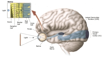
| Retina
-microscopic enlargement
( 200 x )- |
| |
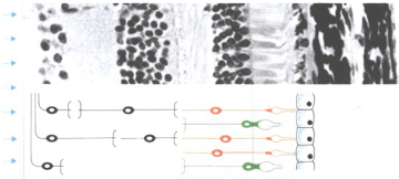 |
|
multipolar
ganglion cells |
|
| the
rod cells |
| bipolar
ganglion cells |
| the cone cells |
|
multipolar
ganglion cells |
|
(light) |
(dark)
pigment cells |
| |
| Light
intensity is perceived with the
rod cells, with the cone
cells |
| colours
can be seen. The fovea
consists of only cone cells,
about |
| 40
000 / mm2. Away from
the fovea gradually the
number of cones |
| are
decreasing and the number of rod
cells are increasing. These |
| parts
of the retina are especially in
use in the twilight when there
is |
| little
light and so colours seem to
have vanished. |
| In
the sense part of the
photoreceptors -the rod cells
and the cone |
| cells-
or photo sensors, a large number
of filmy discs are piled
up |
| perpendicular
to the cell axis. In a cone cell
these discs are about |
| 5
nm thick, in a rod cell about
3.5 nm. On the surface of these
discs |
| lays
the material (a special pigment
called rhodopsin) that is |
| converted
by light (energy) by which the
start of the impulse is |
| produced
necessary to see. The rhodopsin
on the discs in the cone |
| cells
is of three types particularly
sensitive to violet-blue, green and |
|
yellow-orange light.
Each of these rhodopsin forms
absorbs a specific |
| part of the
projected
light by which the action
potential is produced to |
|
perceive colours. |
| |
|
Relation of wavelengths to cone
receptivity: |
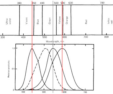 |
| |
| Looking
at these 'figures', for that
matter, they might give you also |
| some
thought why it seems so pleasant
to look into a garden or any |
| landscape
like that. |
| Of
course many things in such a
complex process can go different |
| from
what could be considered as
standard or optimally, due to
the |
| laws
of variation in life or
evolutionary development. That
some |
| of
us actually do see colours
differently has indeed a
relation with |
| the
photosensitivity of the light sensitive
pigments in their cone cells |
| or
even the missing of these
pigments. (Just
click here for some |
| more
information about this subject). |
|